|
|
Volume 37, pages 578-587, 1952 IRIS AGATE FRANCIS T. JONES, 244 Trinity Ave., Berkeley 8, California. ABSTRACT Iris agate owes its spectral colors to the presence of a diffraction grating structure. The lines in the grating are the edges of thin lamellae having alternately high and lower refractive indices. The rhythmic segregation of opal among the crystals may account for the index variation. The chalcedony needles are always perpendicular to the lamellae: The optical properties of the chalcedony associated with the grating structure indicate, that the aggregate is pseudo-orthorhombic, probably being made up of needles of quartz elongated perpendicular to c, but not being perfectly parallel. Form-birefringence may account for the difference of the refractive indices from those of quartz. Maximum and minimum indices are given. Iris agate is agate whose thin section shows spectral colors when viewed in transmitted light. These colors are not thin-film colors such as those of precious opal. Specimens of iris agate were observed in 1933 by Fred S. Young,1,2 then publisher of the Oregon Mineralogist, during the preparation of cabochons. Short articles were published on the subject by H. C. Dake,3,4 editor of the journal. These articles were largely devoted to the occurrence and cutting of such material. Microscopic study by Dr. E. W. Lazell revealed the lamellar structure of iris agate but no detailed study was made of the specimens.1 In 1935 when Fred S. Young and Dr. H. C. Dake first showed the author thin slices of iris or rainbow agate, the display of colors recalled the similar appearance of a diffraction grating. Microscopical examination soon confirmed the opinion that a periodic banded structure existed in the agate, and revealed interesting correlations with the chalcedony's structure and variations in fineness and regularity. No satisfactory explanation of the formation of this regularly banded structure has yet been devised although the theory proposed by Hedges 5 may apply in a general way.OCCURRENCE
It is probable that some specimens of agate from any locality may show the iris structure. Those used in this study were
from Oregon, Montana, and California. Some amygdules from an outcrop near the junction of Grizzly Peak Blvd. and Fish
Ranch Road in Berkeley, Calif., In order to produce the best play of colors the slice of iris agate must he cut so that the thin layers, whose edges are the lines of the diffraction
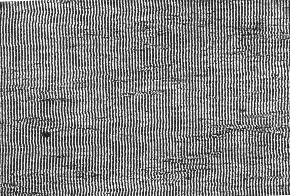
FIG. 1. Iris agate 9,600 lines to the inch. 370X. grating, as shown in Figs. 1 to 5, will be at. right angles to the polished surface. The slice will show the brightest colors if it is thin (1 mm. or less) because the absorbing and scattering effect of any milkiness will then be at a minimum. If the line spacing is uniform, as in Fig. 1, a very thin slice will show primary colors even if the layers in the agate are somewhat inclined to the polished or lacquered surface, although the slice may have to be tilted to correct for the inclination. A specimen which shows primary colors in very thin section may show only pastel colors if it is cut too thick. If the line spacing is not quite uniform only pastel colors may be seen, and if it is very irregular only a chatoyance will be observed. A single band of uniform spacing will show only one color for a given position but any adjacent band of uniform but different spacing will give a different color for the same position because a fine spacing will diffract any one wave length through a larger angle than a coarser grating. In addition, the finer gratings separate the colors from each other more than do the coarser gratings, consequently they give purer, more saturated colors.
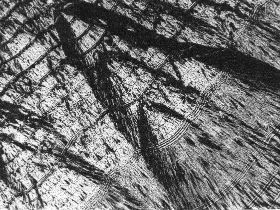
FIG. 2. Iris agate between crossed nicols showing single lines and turtleback. 80X. LINES PER INCH The fineness of structure observed in iris agates ranges from about fifteen thousand to as few as four hundred lines per inch. Figure 1 shows; a specimen having about 10,000 lines per inch. Part of Fig. 2 shows only 400 lines per inch. Finer and coarser spacings may exist in some agates but any spacing in this range will show the grating effect. A grating of only 100 lines per inch will show diffraction colors but they will be so close to the illuminating beam that they will be difficult to see. Gratings as fine as 30,000 lines per inch have been observed by the author in some crystalline organic materials. A variation in spacing of the lines such as shown in Figs. 2 and 3 may possibly be caused by changes in the rate of crystallization of the chalcedony, either because changes in temperature occurred or because the composition (concentration) of the solution (perhaps a gel) varied, or both. It is also possible that the coarse spacing shown in Fig. 2 may be of the ordinary successive deposition type with the grating structure starting, but failing to continue in each of the broad layers which happen to be fairly uniform in thickness. The fact that the needles seem to be continuous across several layers may be explained in the same way phantom lines in single crystals are explained.
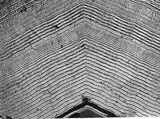
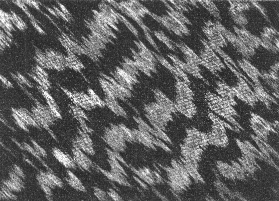
FIG. 3. Iris agate showing spacing differences and change of direction at grain boundary. 80X. CORRELATION OF GRATING LINES AND CRYSTALS The lines in iris agate are always at right angles to the length of the needle-like crystals of chalcedony. Figure 1 shows an unusually straight set of lines but even these are slightly wavy because the crystals are not all exactly parallel to each other. It should be pointed out also that the waves run along the layers within the specimen at right ankles to the cut surface, but the amplitude of the waves is small and a thin slice will give good color in spite of the front to back waviness. Figure 2 shows lines that curve because the crystals have grown in radiating clusters. This structure is sometimes termed "turtleback" or "spherulitic." Figure 3 shows a relatively coarse structure of varying fineness and also reveals that the banding is continuous across adjacent groups of crystals in spite of abrupt changes in direction. Figure 4 is the same specimen as Fig. 1 but seen between crossed nicols and at only 80X. The irregular, ragged black bands indicate changes in polarization colors of the chalcedony but do not correlate with the grating structure. (See Fig. 5.) Such coarse patterns appear to the unaided eye as a woven cloth such as burlap.
FIG. 4. Iris agate between crossed nicols. 80X.
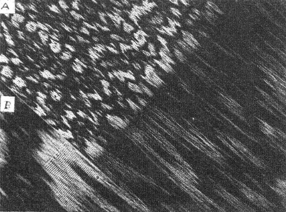
FIG. 5. A. Iris agate between crossed nicols showing start of narrow band where crystal size changes. 80X. B. Start of grating band seen in A. 200X.
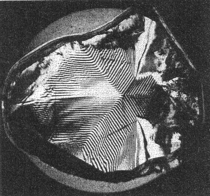
Figure 5A shows a band of iris starting where short, relatively jumbled crystals change to long, nearly parallel crystals. Figure 5B shows the start of the grating band at higher magnification. In most specimens the iris bands show a higher order of polarization color arid more uniform orientation than adjacent bands which do not show the iris grating structure; however, these differences are not always noticeable. The band of iris which begins where the crystal size changes (Fig. 5) becomes finer where a band of cloudy material extends across the field but becomes coarser again beyond the cloudy zone. This cloudy material may be opal which can exist between the crystals of chalcedony. 6 Colloidally dispersed impurities such as the brown ferric hydroxide type of gelatinous appearing "moss" often occur in the bands between the iris bands, but very little such colored impurity is ever present in the iris bands themselves. In any case it appears that the chalcedony in which the grating structure is present must be quite pure and sufficiently free from porosity to prevent much diffusion of iron stain or dyes between the crystals.The observations above indicate that the grating lines must have been formed while the crystals were growing but it is the author's opinion. that the manner of their deposition is not the same as the irregular deposition of the ordinary bands in agate. Organic materials such as cholesteryl acetate melted under a cover glass have been observed crystallizing rapidly in rhythmically banded structures which produce a display of rainbow colors just as does iris agate. Probably a similar process proceeding at a slower rate is responsible for the grating structure in iris agate. NATURE OF THE LINES The lines in iris agate are optical inhomogeneities but are not layers of opaque impurities. If they are impurities they must be of transparent material. The crystals appear to be crossed by grating lines without any disturbance of their optical properties. The lines are visible because an abrupt change in refractive index occurs on both sides of each "line" which must be the edge of a thin layer of material. The boundaries between layers of differing refractive indices cause the black lines in the photomicrographs. The layers of higher index must necessarily alternate with those of lower index. Although a difference of composition could account for such an alternation of refractive index, it is also possible to have such a refractive index difference with constant composition but change in orientation caused by twinning. The grating structure in the amethyst of Fig. 6 is too coarse to show colors but it is conceivable that similar but much finer structures might exist. In this example there are uniform overlapping layers of alternately left- and right-handed quartz. An important difference to be noted is that the grating lines in iris agates go from crystal to crystal whereas the twinning of amethyst is within a single crystal. Zapffe and Worden 7 describe banded structures of somewhat similar appearance revealed by fractography. These structures are confined to single crystals of ammonium dihydrogen phosphate containing some arsenate, but they may have something in common with the lamellar structure in iris agate because an opportunity existed for segregation of arsenate and phosphate in the solid solution which could account for the refractive index differences of the alternate layers. Neither structure has the appearance of twinning.In Figs. 2 and 3 the single thin "lines" or layers have appreciable thickness and the refractive index of each thin layer is greater than the index of the broader crystalline zone on either side. This evidence rules out the possibility that the thin layers contain more opal than the adjacent material. On the contrary they have properties more nearly approaching those of quartz.
FIG. 6. Brazilian twinning in amethyst. (X nicols.) 2X. OPTICAL AND CRYSTALLOGRAPHIC PROPERTIES Table 1 summarizes the optical and crystallographic properties and gives for comparison the corresponding values reported by Dana, 8 Rogers,9 Winchell,10 and Larsen.11Winchell quotes the results of Washburn and Navias 12 who made a careful study of chalcedony from Yellowstone Park and concluded on the basis of x-ray powder diffraction photographs of their sample that chalcedony is quartz in spite of optical, thermal, and density differences. The more recent work of Correns and Nagelschmidt13 indicates that the chalcedony fibers are quartz, elongated at right angles to the c axis. None of
TABLE 1. OPTICAL AND CRYSTALLOGRAPHIC PROPERTIES OF CHALCEDONY AGGREGATES
the authors cited above mention the presence of such a grating structure in agate as is described in this paper. The properties reported here were observed on chalcedony that contained the iris grating structure. Most of the measurements were made on chips broken off the edges of the thin section shown in Fig. 1. Immersion methods were used to observe the edges of the specimen. Sodium light was used for the index determinations which could be made with a precision of ±0.0005, but it should not be expected that all chalcedony specimens would have these values within such narrow limits. The maximum and minimum values observed are given. The black areas in Figs. 4 and 5 reveal crystals oriented in positions which have very low retardation. The centers of these dark areas do note become bright when the specimen is rotated. Interference figures obtained from some of these areas by means of an oil immersion lens, although somewhat indistinct, were good enough to reveal that the crystals, appear to be biaxial. The optic axial angle in oil, 2H=34°; in air-= 2E=50±5°; the optical character is (+) and the axial plane is lengthwise of the crystals, with the acute bisectrix perpendicular to the crystals, The sign of elongation is (-). The bright crystals which show sharp extinction give an "optic normal" interference figure. This combination of properties, if unqualified, would indicate that the crystals, or at least their aggregates, belong in the orthorhombic system, or the monoclinic system if the crystals are elongated in the direction of the b axis. The refractive index for the lengthwise vibrations is always alpha and has a value of 1.5350±0.0005. The refractive index for crosswise vibrations measured on the crystals showing the greatest retardation and sharpest extinction (the "optic normal" view) is gamma and has a value of 1.5430±0.0005. The beta index measured on crystals showing the "acute bisectrix" could not be determined as accurately but has a value of 1.537±0.001. Indices measured on specimens shown in Figs. 2 and 3 show that the single "lines" or layers have a y value of 1.544 compared to the portions of the crystals between the layers which showed a maximum value of 1.543. The change from the "optic normal" to the "acute bisectrix" position occurs quite regularly along any one line of lengthwise (spiral?) growth e.g. Figs. 4 and 5, although this periodicity is not so noticeable in some a specimens, e.g. Fig. 2, where most of the dark crystals are simply near their extinction positions. As noted previously the grating structure does not correlate with this rough periodicity in the variation of orientation. With axial illumination extinction was always parallel with respect to the individual crystals, or small bundles of essentially parallel needles, but in some crystal clusters extinction appeared oblique by about five degrees when observed in highly convergent illumination. These imperfections can be explained if it is recalled that the structure variations are the same perpendicular and horizontal to the cut surface. Any nearly "isotropic" area is composed of a bundle of needle-like crystals which are not perfectly arranged so as to act as a single crystal. The lower layers may contain a few crystals in a position to show the optic normal, others may be dipping slightly. If the needle-like crystals in these specimens of chalcedony were quartz having the c axis lengthwise, then end views of the needles should be "isotropic." Sections cut to show this end view reveal that the crystals are not "isotropic" but show a retardation about equal to that seen on sections cut parallel to the length of the crystals. The thin layers responsible for the grating structure do not show any variation in their extinction position as do twins, and as mentioned earlier, these lamellae have refractive indices greater than the zones between them. The maximum observed difference is only 0.001, but the contrast within the specimen would lead one to estimate a larger value. DISCUSSION On the basis of the evidence given above one would conclude that: (1) The iris agate grating structure results from the segregation of alternate layers having higher and lower refractive indices during crystallization of the needles or fibers of chalcedony. (2) The crystallization is a rhythmic process affecting the entire aggregate of crystals uniformly. (3) Factors such as variations in rate of growth and percentage of impurity, such as opal, probably cause the difference of fineness in the different grating bands of a specimen. If Donnay's6 explanation of the influence of form-birefringence is accepted, the higher index of the thin layers in. some specimens such as Figs. 2 and 3 cannot be attributed to an increased amount of opal but the lower index of adjacent material may be accounted for on that basis. The optical properties of the aggregate would indicate that chalcedony is not quartz but an orthorhombic or monoclinic mineral. However, if allowance is made for the fact that these properties are measured on an aggregate of very small crystals instead of upon single crystals, and also considering the influence of amorphous intercrystalline material, probably opal, upon the index values6 it is conceivable that the difference between these optical properties and those of quartz (e =1.553, m =1.544) can be accounted for. It is also necessary to explain the difference in habit of the chalcedony needles (c axis perpendicular to length) in contrast with that of ordinary quartz. It is to be noted that both quartz and chalcedony are optically positive. Unless there is some ambiguity l4 in the x-ray data or possibly very slight but significant differences between the diffraction patterns of chalcedony and quartz, chalcedony crystals must be considered quartz in an unusual form. An x-ray fiber pattern obtained from a sliver of iris agate gave dimensions agreeing with those measured on a powder pattern of alpha quartz.Manuscript received July 27, 1951
NOTES 1Young, Fred S., Private communication. 2 Young, Fred S., Oregon Mineralogist, 2, 22 (April 1934). 3 Dake, H. C., Oregon Mineralogist, 1, 1 (July 1933). 4 Dake, Fleener and Wilson, Quartz Family Minerals, McGraw-Hill Book Co., (1938). 5 Hedges, E. S., Liesegang Rings and
Other Periodic Structures, London, Chapman & Hall, Ltd. (1932). 6 Donnay, J. D. H., Le birefringence de forme Bans la calcedoine: Ann. Soc. Geol. Belgique, 59, B 289 (1936). 7 Zapffe and Worden, Acta Crystallographica, 2, 386 (1949). 8 Dana and Ford, Textbook of Mineralogy, 4th Ed., John Wiley and Sons, Inc., (1932).9 Rogers, Introduction to the Study of Minerals; 3rd Ed., McGraw-Hill Book Co. (1937).10 Winchell, Microscopic Characters of Artificial Minerals, John Wiley and Sons, Inc (1931). 11 Larsen and Berman, The Microscopic Determination of the Nonopaque Minerals U.S.G.S., Bulletin 848 (1934).12 Washburn and Navias, Proc. Nat'l. Acad. Sci., 8, 1 (1922). 13 Correns and Nagelschmidt, Zeits. Krist., 85, 199 (1933). 14 Menzer, G. Z., Naturforsch: 4a, 11-21 (April, 1949). (Only the abstract seen.) [var:'startyear'='1952'] [Include:'footer.htm'] |
